Diamond-Blackfan anemia (DBA) is a hereditary bone marrow failure disorder that is characterized by erythropoiesis failure and chronic anemia and is frequently associated with physical malformations. Considered a ribosomopathy, most patients harbor pathogenic mutations in one of at least 22 large or small ribosomal protein genes in an autosomal dominant manner, however rarer cases involving mutations in GATA1, TSR2 and EPO have also been described.
A recent study of the architecture of the human hematopoietic hierarchy throughout development (Notta et al, Science 2016) showed that, after birth, unipotent progenitors can derive directly from multipotent progenitors without intermediate differentiation into oligopotent progenitors. Critically, this work identified novel progenitors for which the abundance, clonal capacity and progenitor function is currently unknown in many benign and malignant hematological disorders. Specifically, multipotent, common myeloid and megakaryocyte-erythroid progenitors (MPP, CMP and MEP) were found to be functionally heterogeneous and could be subdivided (F1, F2 and F3 subtypes) based on the combination of CD71 and BAH1 expression. We hypothesized that in-depth analysis of these novel and previously reported progenitors will better elucidate hematopoiesis in DBA, providing insights in to DBA pathogenesis and erythropoietic failure.
Using flow cytometry, we quantified the frequencies of 11 hematopoietic stem and progenitor cell (HSPC) populations: HSC, MPP F1-F3, CMP F1-F3, MEP F1-F3 and granulocyte-monocyte progenitors (GMP), from the BM specimens of 8 DBA patients harboring ribosomal protein mutations and 6 healthy age-matched control donors. To characterize the function of HSPCs in DBA we then utilized a highly sensitive and quantitative single-cell in-vitro stromal feeder-based differentiation assay that supports the expansion and lineage commitment of single HSCPs towards mature cell types (myeloid, erythroid and megakaryocytes) with progeny immunophenotyping and quantification by flow cytometry.
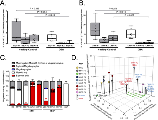
Our refined quantification of HSPCs by flow cytometry showed that the defect of erythropoiesis in DBA patients starts at the CMP/MEP compartment. Namely, CMP-F2 and CMP-F3 as well as MEP-F2 and MEP-F3 sub-populations that are predicted to form erythroid cells were significantly reduced in DBA patients compared to healthy aged-matched individuals (Figure 1A-B). In contrast, CMP/MEP-F1 sub-populations that are predicted to form only myeloid cells (BAH1-/CD71-) showed no significant difference compared to healthy controls. Importantly, the multipotent compartment (HSC and MPP) did not significantly differ from that of healthy controls. Functional analysis of F2/F3 CMP/MEP HSCP sub-populations in single-cell colony assays showed that they have a high propensity to produce erythroid progeny in healthy control samples (Figure 1C). In contrast, in DBA patients these HSCPs had limited erythroid forming potential, but maintain a high propensity to develop myeloid colonies (Figure 1D).
To our knowledge this is the first study that the HSCP sub-populations F1/F2/F3 HSCP fractions have been analyzed in DBA. Collectively, these data indicate that the hematopoietic defect in DBA occurs in erythroid-producing CMP/MEP populations. Our data show that these sub-populations are both reduced and dysfunctional in DBA patients and we suggest these abnormalities lead to the pure red cell aplasia seen in these patients.
Figure 1: DBA patients have reduced CMP and MEP sub-populations that behave dysfunctional in-vitro. A-B) DBA patients have reduced CMP-F2/3 (A) and MEP-F2/3 (B) sub-populations in their bone marrow. C) CMP F2-F3 and MEP F2-F3 have a high propensity to produce erythroid cell types in healthy controls. D) Single-cells isolated from the CMP-F2/3 and MEP-F2 population of DBA patients produced proportionally fewer erythroid and greater myeloid colonies in in-vitro colony assays (plot shows the mean respective proportions from all healthy controls and DBA patients tested, each N=5).
Dick:Bristol-Myers Squibb/Celgene: Research Funding.
Author notes
Asterisk with author names denotes non-ASH members.


This feature is available to Subscribers Only
Sign In or Create an Account Close Modal